
Caring for a horse involves paying attention to many details. Feeding routines, training demands, turnout opportunities, and daily habits all shape how a horse feels and behaves. Owners often notice when something seems slightly off, even if the change is small.
When a horse becomes less willing under saddle, shows changes in appetite, or isn’t performing quite the same, gastric ulcers become part of the discussion. Ulcers are common in horses in regular work, especially those exposed to training stress, changes in schedule, or periods without steady access to forage. Because of this, gastric ulcers are often considered when a horse develops unexplained changes.
The important step is determining whether ulcers are present and how significant they are. Gastroscopy for horses allows us to answer that directly.
The Equine Stomach and Why Ulcers Develop
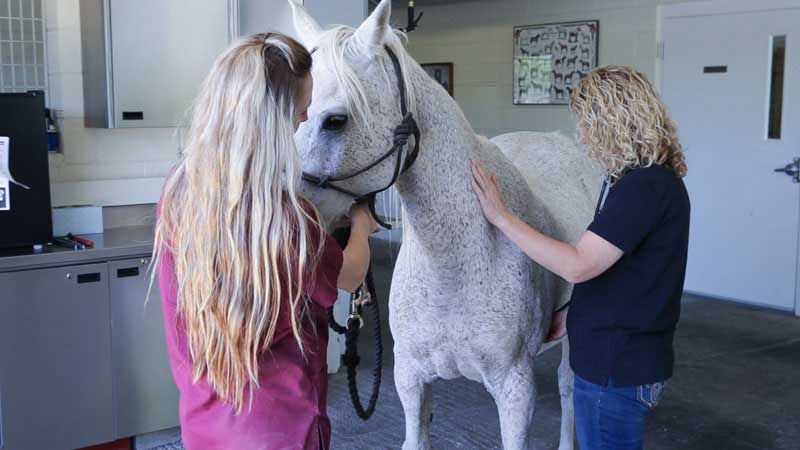
The equine stomach is small relative to body size and built for near-continuous intake of forage. Horses produce gastric acid constantly, not just during meals. Under natural grazing conditions, that steady acid production is buffered by saliva and the continual presence of roughage.
The stomach is one compartment, but it has two distinct regions: the squamous (non-glandular) region at the top and the glandular region at the bottom. Food enters the stomach from the esophagus and passes through the squamous region first before settling into the glandular portion. From there, it moves through the pylorus into the small intestine.
These regions behave differently. The squamous mucosa has limited protection from acid and is especially vulnerable when the stomach is relatively empty or during exercise, when acid can move upward along the stomach wall. The glandular region produces acid but also maintains protective mucus and bicarbonate layers. Ulcers in this area tend to occur when those defenses are disrupted.
Modern management can influence both regions. Meal feeding, high-concentrate diets, long gaps without forage, transportation, confinement, and intense exercise all alter gastric pH and mucosal protection. These differences matter, because treatment decisions depend on which region is affected and how extensive the lesions are.
The Value of Direct Visualization
Because gastric ulcers are common, they are frequently suspected when a horse shows changes in attitude, appetite, or performance. But not every horse with these signs has ulcer disease, and the severity varies widely among those who do.
Gastroscopy for horses remains the most reliable way to diagnose gastric ulcers. Direct visualization allows us to determine whether ulcers are present, where they are located, and how deep or extensive they are. Some horses have small, superficial lesions. Others show significant glandular disease. These distinctions influence medication choices, duration of therapy, and follow-up recommendations.
Gastroscopy also helps assess healing. Improvement in behavior or appetite alone does not always reflect complete resolution.
What Gastroscopy Involves
Gastroscopy for horses is performed using a three-meter flexible video endoscope. The horse remains standing under light sedation, and the procedure is not surgical.
Feed is withheld for approximately 12 hours to allow a clear view of the stomach lining. Water is restricted several hours before the exam.
The endoscope is passed through the nostril, down the esophagus, and into the stomach. We examine the esophagus, the squamous region, the glandular region, and the pyloric outflow in a systematic manner. Once the stomach is reached, the examination typically takes 15 to 20 minutes.
Most horses tolerate the procedure well and return to normal activity the same day.
Why Gastroscopy Is Recommended
Gastroscopy is recommended when a horse’s performance declines without an obvious orthopedic reason, when recurrent mild colic occurs, when appetite or body condition changes are noted, or when previous ulcer treatment has not resolved concerns. It is also useful when confirmation of healing is needed before returning to full work.
Gastroscopy is the only reliable way to confirm gastric ulcers and evaluate their severity.
If you would like to see a brief demonstration of what a gastroscopy looks like, this short educational video offers a simple overview: Equine Gastroscopy 101.
What the Results Mean for Your Horse
Equine gastric ulcer disease is common, but each case is different. Management practices, stress level, exercise intensity, and individual sensitivity all contribute to the development and severity of disease.
Gastroscopy for horses provides clear, objective findings. It allows treatment and management recommendations to be based on documented evidence rather than assumptions.
If your horse is showing changes in performance, appetite, or behavior that raise concern for gastric ulcers, a gastroscopy examination can help determine what is truly happening and what the next steps should be.
When the answers are not obvious, Adobe Veterinary Center is here to help. Our Equine Internal Medicine service, led by Dr. Laura Weintraub, provides advanced diagnostics for horses with complex or unexplained health concerns. Dr. Weintraub is the only equine internal medicine specialist in Tucson, allowing horse owners in the region to access specialized care without the stress of a multi-hour haul.
If you are concerned about your horse’s gastric health or would like to discuss whether gastroscopy may be appropriate, contact Adobe Veterinary Center to schedule an examination or consultation.